01 PLANES & LEVELS
I have compiled for you all the key landmarks and their levels that you need to learn. Download the PDF and give yourself 5 minutes to memorise! It is super high-yield for both Part A and Part B.
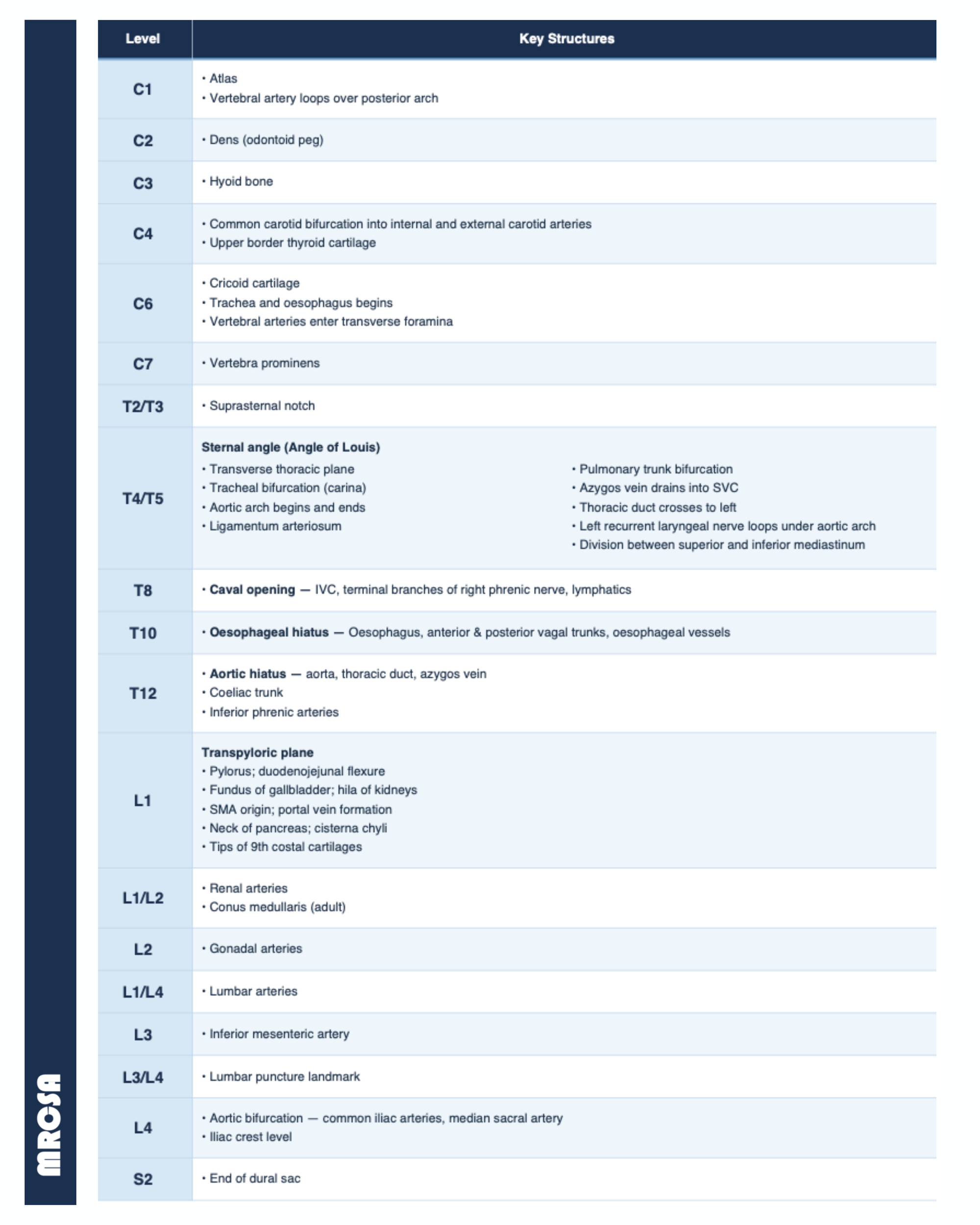
x4 bifurcations to memorise:
C4 - Common carotid artery bifurcates into the external and internal carotid arteries; this is also the level of the upper border of the thyroid cartilage.
T4/T5 - Sternal Angle of Louis - Trachea bifurcates at the carina into the right and left main bronchi; the right bronchus is wider, shorter and more vertical.
L4 - Abdominal aorta bifurcates at the level of the umbilicus into the left and right common iliac arteries.
L5 - Inferior vena cava is formed by the union of the left and right common iliac veins.
👩⚕️ BiFOURcations at C4, T4/5 and L4
x3 diaphragmatic openings to memorise:
T8 - Caval hiatus: Transmits the IVC and right phrenic nerve.
T10 - Oesophageal hiatus: Transmits the oesophagus, vagal anterior and posterior trunks, and oesophageal branches.
T12 - Aortic hiatus: Transmits the aorta, thoracic duct, azygos vein, sometimes hemiazygos vein.
👩⚕️ Vena cava, 8 letters, at T8; Oesophagus, 10 letters, at T10; Aortic hiatus, 12 letters, at T12.
Finally, these two planes are high yield:
Angle of Louis at T4/T5
- Bifurcation of trachea (carina)
- Arch of aorta begins and ends
- Ligamentum arteriosum
- Pulmonary trunk bifurcation just below, at T5
- Azygous vein drains into SVC
- Thoracic duct crosses to left side
- Left recurrent laryngeal nerve hooks around arch (near ligamentum arteriosum)
- Marks the division between superior and inferior mediastinum
- Level of thoracic spine T4/T5 disc posteriorly
Transpyloric Plane at L1
- Pylorus of stomach (hence the name)
- Duodenojejunal flexure - to the left at the transpyloric plane
- Fundus of gallbladder
- Hila of kidneys
- Origin of superior mesenteric artery SMA
- Portal vein formation behind neck of pancreas
- Pancreatic neck & body
- Cisterna chyli
- Tips of 9th costal cartilages
- Conus medullaris (end of spinal cord in adults)

Which of the following structures passes through the diaphragmatic opening at T12?
- ((Vagus nerve and oesophagus::Enter at T10))
- ((IVC and phrenic nerve::Enter at T8))
- ((Ureter and gonadal vessels::Don't pass through a diaphragmatic opening))
- ((Azygos vein and thoracic duct::☑️ The aorta, thoracic duct, azygos vein and sometimes hemiazygos pass through the diaphragmatic opening at T12))
The inferior vena cava passes through the diaphragm at which vertebral level?
- ((T8::☑️ Caval opening; inferior vena cava passes through the diaphragm at T8))
- ((T9::No major diaphragmatic aperture))
- ((T10::Oesophageal hiatus — oesophagus and vagal trunks))
- ((T11::No major diaphragmatic aperture))
Which of these do not occur at the transpyloric plane?
- ((Fundus of the gallbladder::The gallbladder fundus lies at the transpyloric plane, typically at the tip of the 9th costal cartilage in the mid-clavicular line))
- ((Neck of the pancreas::The neck of the pancreas sits directly on the transpyloric plane at L1))
- ((Hilum of the kidneys::Both renal hila lie at the transpyloric plane: the left at L1, the right slightly lower but still within the plane.))
- ((None of the above::☑️ Correct, all of the listed structures are located at the transpyloric plane))
At which level is the neck of the pancreas?
- ((T12::Level of the aortic hiatus))
- ((L1::☑️ The neck of the pancreas lies on the transpyloric plane at L1))
- ((L2::Below the level of the neck of the pancreas))
- ((L3::Too low — near the inferior border of the pancreas))
A structure located at the level of the T5 vertebra is:
- ((Left atrium::Incorrect))
- ((Tracheal bifurcation::Incorrect))
- ((Bifurcation of pulmonary trunk::☑️ See how the pulmonary trunk bifurcates at T5, inferior to the tracheal bifurcation at T4/T5))
- ((Bifurcation of right bronchus::Incorrect))